
Page 298 - Atlas of Histology with Functional Correlations
P. 298
anchors them to the Z line, and functions in regulating the length of the actin
filament. Further support is provided by the protein desmin that extends from Z
line of one myofibril to the adjacent myofibril, linking them together and
attaching them to the sarcolemma (cell membrane). This stabilizes the position
of muscle myofibrils within the sarcoplasm.
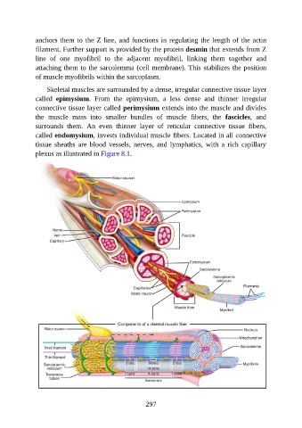
Skeletal muscles are surrounded by a dense, irregular connective tissue layer
called epimysium. From the epimysium, a less dense and thinner irregular
connective tissue layer called perimysium extends into the muscle and divides
the muscle mass into smaller bundles of muscle fibers, the fascicles, and
surrounds them. An even thinner layer of reticular connective tissue fibers,
called endomysium, invests individual muscle fibers. Located in all connective
tissue sheaths are blood vessels, nerves, and lymphatics, with a rich capillary
plexus as illustrated in Figure 8.1.
297