
Page 301 - Atlas of Histology with Functional Correlations
P. 301
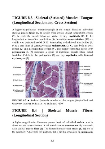
FIGURE 8.3 | Skeletal (Striated) Muscles: Tongue

               (Longitudinal Section and Cross Section)


               A  higher-magnification  photomicrograph  of  the  tongue  illustrates  individual
               skeletal muscle fibers (3, 9) in both cross section (3) and longitudinal section

               (9).  In  each,  the  muscle  fibers  are  visible  as  tiny  myofibrils  (4).  In  the
               longitudinal section of the muscle fiber (9), the multiple cross-striations (10) are
               visible with peripheral nuclei (5, 9). Surrounding each skeletal muscle fiber (3,

               9) is a thin layer of connective tissue endomysium (2, 6),  seen  both  in  cross
               section (2) and in longitudinal section (6). The thicker connective tissue layer
               perimysium  (1,  7)  surrounds  a  group  of  individual  muscle  fibers  called
               fascicles.  Visible  in  the  perimysium  (7)  are  tiny  capillaries  with  flattened
               erythrocytes (8).


               FIGURE  8.3  ■  Skeletal  (striated)  muscles  of  the  tongue  (longitudinal  and
               transverse section). Stain: Masson trichrome. ×130.


               FIGURE                8.4         |      Skeletal            Muscle             Fibers

               (Longitudinal Section)


               A  higher-magnification  illustrates  greater  detail  of  individual  skeletal  muscle
               fibers and the cross-striations. A cell membrane, or sarcolemma (4), surrounds
               each skeletal muscle fiber (2). The flattened muscle fiber nuclei (1, 10) are in
               the periphery. Adjacent to the nuclei (1, 10) is the thin cytoplasm or sarcoplasm


                                                          300