
Page 302 - Atlas of Histology with Functional Correlations
P. 302
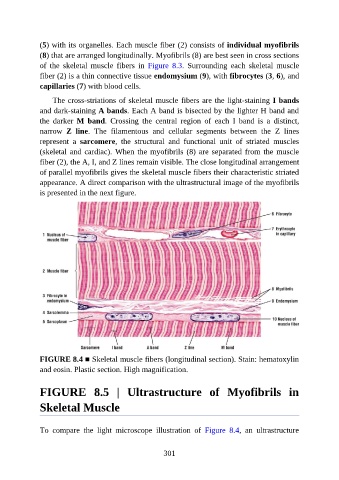
(5) with its organelles. Each muscle fiber (2) consists of individual myofibrils

               (8) that are arranged longitudinally. Myofibrils (8) are best seen in cross sections
               of  the  skeletal  muscle  fibers  in  Figure  8.3.  Surrounding  each  skeletal  muscle
               fiber (2) is a thin connective tissue endomysium (9), with fibrocytes (3, 6), and
               capillaries (7) with blood cells.

                   The cross-striations of skeletal muscle fibers are the light-staining I bands

               and dark-staining A bands. Each A band is bisected by the lighter H band and
               the  darker  M  band.  Crossing  the  central  region  of  each  I  band  is  a  distinct,
               narrow  Z  line.  The  filamentous  and  cellular  segments  between  the  Z  lines
               represent  a  sarcomere,  the  structural  and  functional  unit  of  striated  muscles

               (skeletal and cardiac). When the myofibrils (8) are separated from the muscle
               fiber (2), the A, I, and Z lines remain visible. The close longitudinal arrangement
               of parallel myofibrils gives the skeletal muscle fibers their characteristic striated
               appearance. A direct comparison with the ultrastructural image of the myofibrils

               is presented in the next figure.


               FIGURE 8.4 ■ Skeletal muscle fibers (longitudinal section). Stain: hematoxylin
               and eosin. Plastic section. High magnification.


               FIGURE  8.5  |  Ultrastructure  of  Myofibrils  in

               Skeletal Muscle


               To  compare  the  light  microscope  illustration  of  Figure  8.4,  an  ultrastructure


                                                          301