
Page 307 - Atlas of Histology with Functional Correlations
P. 307
and activate complex reflexes to regulate muscle activity. When skeletal
muscles are stretched, the neuromuscular spindles initiate a reflex contraction
and shortening of the muscle.
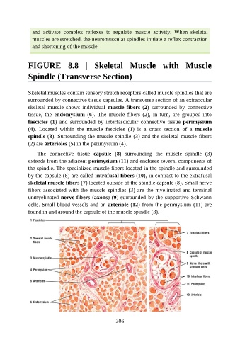
FIGURE 8.8 | Skeletal Muscle with Muscle
Spindle (Transverse Section)
Skeletal muscles contain sensory stretch receptors called muscle spindles that are
surrounded by connective tissue capsules. A transverse section of an extraocular
skeletal muscle shows individual muscle fibers (2) surrounded by connective
tissue, the endomysium (6). The muscle fibers (2), in turn, are grouped into
fascicles (1) and surrounded by interfascicular connective tissue perimysium
(4). Located within the muscle fascicles (1) is a cross section of a muscle
spindle (3). Surrounding the muscle spindle (3) and the skeletal muscle fibers
(2) are arterioles (5) in the perimysium (4).
The connective tissue capsule (8) surrounding the muscle spindle (3)
extends from the adjacent perimysium (11) and encloses several components of
the spindle. The specialized muscle fibers located in the spindle and surrounded
by the capsule (8) are called intrafusal fibers (10), in contrast to the extrafusal
skeletal muscle fibers (7) located outside of the spindle capsule (8). Small nerve
fibers associated with the muscle spindles (3) are the myelinated and terminal
unmyelinated nerve fibers (axons) (9) surrounded by the supportive Schwann
cells. Small blood vessels and an arteriole (12) from the perimysium (11) are
found in and around the capsule of the muscle spindle (3).
306