
Page 311 - Atlas of Histology with Functional Correlations
P. 311
much change in their diameters. Also, cardiac muscle fibers are shorter than a
skeletal muscle fiber with a single, centrally located nucleus (3, 7). Binucleate
(two nuclei) muscle fibers (8) are also occasionally seen. The nuclei (7) are
visible in the center of each muscle fiber when cut in a transverse section.
Around these central nuclei (3, 7, 8) are the clear zones of nonfibrillar
perinuclear sarcoplasm (1, 13). In transverse sections, the perinuclear
sarcoplasm (13) appears as a clear space if the section is not through the nucleus.
Also visible in transverse sections are individual myofibrils (14) of cardiac
muscle fibers.
Highly characteristic features of cardiac muscle fibers are the intercalated
discs (4, 9). These dark-staining structures are found at irregular intervals and
represent the specialized junctional complexes between cardiac muscle fibers.
Cardiac muscle has a vast blood supply. Numerous small blood vessels and
capillaries (6) are found in the connective tissue (11) septa and the delicate,
indistinct endomysium (12) between individual muscle fibers.
Other examples of cardiac muscles are seen in Chapter 10.
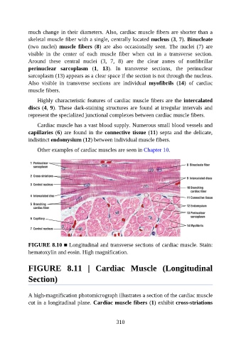
FIGURE 8.10 ■ Longitudinal and transverse sections of cardiac muscle. Stain:
hematoxylin and eosin. High magnification.
FIGURE 8.11 | Cardiac Muscle (Longitudinal
Section)
A high-magnification photomicrograph illustrates a section of the cardiac muscle
cut in a longitudinal plane. Cardiac muscle fibers (1) exhibit cross-striations
310