
Page 313 - Atlas of Histology with Functional Correlations
P. 313
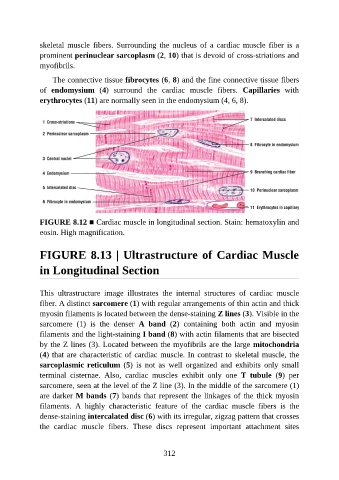
skeletal  muscle  fibers.  Surrounding  the  nucleus  of  a  cardiac  muscle  fiber  is  a

               prominent perinuclear sarcoplasm (2, 10) that is devoid of cross-striations and
               myofibrils.

                   The connective tissue fibrocytes (6, 8) and the fine connective tissue fibers
               of  endomysium  (4)  surround  the  cardiac  muscle  fibers.  Capillaries  with
               erythrocytes (11) are normally seen in the endomysium (4, 6, 8).


               FIGURE 8.12 ■ Cardiac muscle in longitudinal section. Stain: hematoxylin and
               eosin. High magnification.


               FIGURE 8.13 | Ultrastructure of Cardiac Muscle

               in Longitudinal Section


               This  ultrastructure  image  illustrates  the  internal  structures  of  cardiac  muscle
               fiber. A distinct sarcomere (1) with regular arrangements of thin actin and thick
               myosin filaments is located between the dense-staining Z lines (3). Visible in the
               sarcomere  (1)  is  the  denser  A  band  (2)  containing  both  actin  and  myosin

               filaments and the light-staining I band (8) with actin filaments that are bisected
               by the Z lines (3). Located between the myofibrils are the large mitochondria
               (4) that are characteristic of cardiac muscle. In contrast to skeletal muscle, the
               sarcoplasmic reticulum (5)  is  not  as  well  organized  and  exhibits  only  small

               terminal  cisternae.  Also,  cardiac  muscles  exhibit  only  one  T  tubule  (9)  per
               sarcomere, seen at the level of the Z line (3). In the middle of the sarcomere (1)
               are darker M bands (7) bands that represent the linkages of the thick myosin
               filaments.  A  highly  characteristic  feature  of  the  cardiac  muscle  fibers  is  the

               dense-staining intercalated disc (6) with its irregular, zigzag pattern that crosses
               the  cardiac  muscle  fibers.  These  discs  represent  important  attachment  sites


                                                          312