
Page 312 - Atlas of Histology with Functional Correlations
P. 312
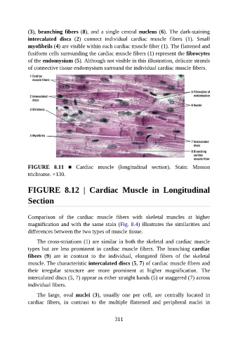
(3), branching fibers (8), and  a  single  central  nucleus (6).  The  dark-staining

               intercalated  discs  (2)  connect  individual  cardiac  muscle  fibers  (1).  Small
               myofibrils (4) are visible within each cardiac muscle fiber (1). The flattened and
               fusiform cells surrounding the cardiac muscle fibers (1) represent the fibrocytes
               of the endomysium (5). Although not visible in this illustration, delicate strands
               of connective tissue endomysium surround the individual cardiac muscle fibers.


               FIGURE  8.11  ■  Cardiac  muscle  (longitudinal  section).  Stain:  Masson

               trichrome. ×130.


               FIGURE  8.12  |  Cardiac  Muscle  in  Longitudinal


               Section


               Comparison  of  the  cardiac  muscle  fibers  with  skeletal  muscles  at  higher
               magnification and with the same stain (Fig. 8.4) illustrates the similarities and
               differences between the two types of muscle tissue.

                   The cross-striations (1) are similar in both the skeletal and cardiac muscle

               types  but  are  less  prominent  in  cardiac  muscle  fibers.  The  branching  cardiac
               fibers  (9)  are  in  contrast  to  the  individual,  elongated  fibers  of  the  skeletal
               muscle. The characteristic intercalated discs (5, 7) of cardiac muscle fibers and

               their  irregular  structure  are  more  prominent  at  higher  magnification.  The
               intercalated discs (5, 7) appear as either straight bands (5) or staggered (7) across
               individual fibers.

                   The  large,  oval  nuclei  (3),  usually  one  per  cell,  are  centrally  located  in
               cardiac  fibers,  in  contrast  to  the  multiple  flattened  and  peripheral  nuclei  in


                                                          311