
Page 308 - Atlas of Histology with Functional Correlations
P. 308
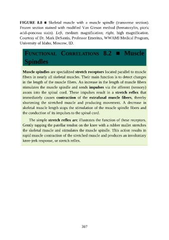
FIGURE  8.8  ■  Skeletal  muscle  with  a  muscle  spindle  (transverse  section).

               Frozen section stained with modified Van Gieson method (hematoxylin, picric
               acid–ponceau  stain).  Left,  medium  magnification;  right,  high  magnification.
               Courtesy of Dr. Mark DeSantis, Professor Emeritus, WWAMI Medical Program,
               University of Idaho, Moscow, ID.


                  FUNCTIONAL  CORRELATIONS  8.2  ■  Muscle


                  Spindles


                 Muscle spindles are specialized stretch receptors located parallel to muscle
                 fibers in nearly all skeletal muscles. Their main function is to detect changes
                 in the length of the muscle fibers. An increase in the length of muscle fibers

                 stimulates the muscle spindle and sends impulses via the afferent (sensory)
                 axons  into  the  spinal  cord.  These  impulses  result  in  a  stretch  reflex  that
                 immediately  causes  contraction  of  the  extrafusal  muscle  fibers,  thereby
                 shortening  the  stretched  muscle  and  producing  movement.  A  decrease  in

                 skeletal muscle length stops the stimulation of the muscle spindle fibers and
                 the conduction of its impulses to the spinal cord.

                     The simple stretch reflex arc illustrates the function of these receptors.
                 Gently tapping the patellar tendon on the knee with a rubber mallet stretches
                 the skeletal muscle and stimulates the muscle spindle. This action results in

                 rapid muscle contraction of the stretched muscle and produces an involuntary
                 knee-jerk response, or stretch reflex.


                                                          307