
Page 304 - Atlas of Histology with Functional Correlations
P. 304
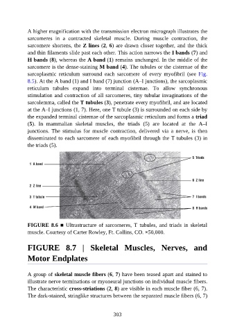
A higher magnification with the transmission electron micrograph illustrates the

               sarcomeres  in  a  contracted  skeletal  muscle.  During  muscle  contraction,  the
               sarcomere shortens, the Z lines (2, 6) are drawn closer together, and the thick
               and thin filaments slide past each other. This action narrows the I bands (7) and
               H bands (8), whereas the A band (1) remains unchanged. In the middle of the
               sarcomere is the dense-staining M band (4). The tubules or the cisternae of the

               sarcoplasmic  reticulum  surround  each  sarcomere  of  every  myofibril  (see  Fig.
               8.5). At the A band (1) and I band (7) junction (A–I junctions), the sarcoplasmic
               reticulum  tubules  expand  into  terminal  cisternae.  To  allow  synchronous

               stimulation and contraction of all sarcomeres, tiny tubular invaginations of the
               sarcolemma, called the T tubules (3), penetrate every myofibril, and are located
               at the A–I junctions (1, 7). Here, one T tubule (3) is surrounded on each side by
               the expanded terminal cisternae of the sarcoplasmic reticulum and forms a triad
               (5).  In  mammalian  skeletal  muscles,  the  triads  (5)  are  located  at  the  A–I

               junctions.  The  stimulus  for  muscle  contraction,  delivered  via  a  nerve,  is  then
               disseminated to each sarcomere of each myofibril through the T tubules (3) in
               the triads (5).


               FIGURE 8.6 ■  Ultrastructure  of  sarcomeres,  T  tubules,  and  triads  in  skeletal

               muscle. Courtesy of Carter Rowley, Ft. Collins, CO. ×50,000.


               FIGURE  8.7  |  Skeletal  Muscles,  Nerves,  and


               Motor Endplates


               A group of skeletal muscle fibers (6, 7) have been teased apart and stained to
               illustrate nerve terminations or myoneural junctions on individual muscle fibers.
               The characteristic cross-striations (2, 8) are visible in each muscle fiber (6, 7).

               The dark-stained, stringlike structures between the separated muscle fibers (6, 7)


                                                          303