
Page 305 - Atlas of Histology with Functional Correlations
P. 305
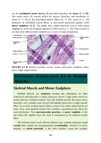
are the myelinated motor nerves (3) and their branches, the axons (1, 5, 10).

               The  motor  nerve  (3)  courses  within  the  muscle,  branches,  and  distributes  its
               axons  (1,  5,  10)  to  the  individual  muscle  fibers  (6,  7).  The  axons  (1,  5,  10)
               terminate  on  individual  muscle  fibers  as  specialized  junctional  regions  called
               motor endplates (4, 9). The small, dark, round structures seen in each motor
               endplate (4, 9) are the terminal expansion of the axons (1, 5, 10). Some axons (1)

               are also seen without motor endplates as a result of tissue preparation.


               FIGURE  8.7  ■  Skeletal  muscles,  nerves,  axons,  and  motor  endplates.  Stain:
               silver. High magnification.


                  FUNCTIONAL  CORRELATIONS  8.1  ■  Skeletal


                  Muscles


                 Skeletal Muscle and Motor Endplates


                     Skeletal  muscles  are  voluntary  because  the  stimulation  for  their
                 contraction and relaxation is under conscious control. Large motor nerves or

                 axons innervate skeletal muscles. Near the skeletal muscle, the motor nerve
                 branches, and a smaller axon branch individually innervates a single muscle
                 fiber. As a result, skeletal muscle fibers contract only when stimulated by an

                 axon. Also, each skeletal muscle fiber exhibits a specialized site where the
                 axon terminates. This neuromuscular junction, or motor endplate, is the
                 site where the impulse from the axon is transmitted to the skeletal muscle
                 fiber.

                     The terminal end of each efferent (motor) axon contains numerous small

                 vesicles that contain the neurotransmitter acetylcholine. Arrival of a nerve
                 impulse,  or  action  potential,  at  the  axon  terminal  causes  the  synaptic


                                                          304