
Page 303 - Atlas of Histology with Functional Correlations
P. 303
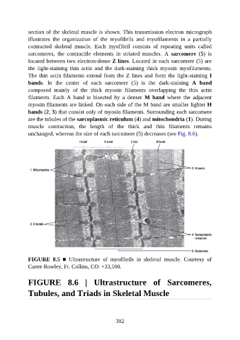
section of the skeletal muscle is shown. This transmission electron micrograph

               illustrates  the  organization  of  the  myofibrils  and  myofilaments  in  a  partially
               contracted  skeletal  muscle.  Each  myofibril  consists  of  repeating  units  called
               sarcomeres,  the  contractile  elements  in  striated  muscles.  A  sarcomere  (5)  is
               located between two electron-dense Z lines. Located in each sarcomere (5) are
               the  light-staining  thin  actin  and  the  dark-staining  thick  myosin  myofilaments.

               The thin actin filaments extend from the Z lines and form the light-staining I
               bands.  In  the  center  of  each  sarcomere  (5)  is  the  dark-staining  A  band
               composed  mainly  of  the  thick  myosin  filaments  overlapping  the  thin  actin

               filaments.  Each  A  band  is  bisected  by  a  denser  M  band  where  the  adjacent
               myosin filaments are linked. On each side of the M band are smaller lighter H
               bands (2, 3) that consist only of myosin filaments. Surrounding each sarcomere
               are the tubules of the sarcoplasmic reticulum (4) and mitochondria (1). During
               muscle  contraction,  the  length  of  the  thick  and  thin  filaments  remains

               unchanged, whereas the size of each sarcomere (5) decreases (see Fig. 8.6).


               FIGURE  8.5  ■  Ultrastructure  of  myofibrils  in  skeletal  muscle.  Courtesy  of
               Carter Rowley, Ft. Collins, CO. ×33,500.


               FIGURE  8.6  |  Ultrastructure  of  Sarcomeres,


               Tubules, and Triads in Skeletal Muscle


                                                          302