Page 299 - Atlas of Histology with Functional Correlations
P. 299
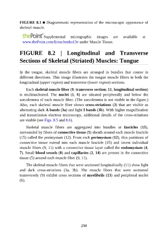
FIGURE 8.1 ■ Diagrammatic representation of the microscopic appearance of

               skeletal muscle.


                                Supplemental       micrographic       images     are    available     at
                 www.thePoint.com/Eroschenko13e under Muscle Tissue.


               FIGURE  8.2  |  Longitudinal  and  Transverse


               Sections of Skeletal (Striated) Muscles: Tongue


               In  the  tongue,  skeletal  muscle  fibers  are  arranged  in  bundles  that  course  in
               different directions. This image illustrates the tongue muscle fibers in both the

               longitudinal (upper region) and transverse (lower region) sections.

                   Each skeletal muscle fiber (9, transverse section; 11, longitudinal section)
               is  multinucleated.  The  nuclei  (1,  6)  are  situated  peripherally  and  below  the
               sarcolemma of each muscle fiber. (The sarcolemma is not visible in the figure.)

               Also, each skeletal muscle fiber shows cross-striations (3)  that  are  visible  as
               alternating dark A bands (3a) and light I bands (3b). With higher magnification
               and transmission electron microscopy, additional details of the cross-striations
               are visible (see Figs. 8.5 and 8.6).

                   Skeletal  muscle  fibers  are  aggregated  into  bundles  or  fascicles  (15),

               surrounded by fibers of connective tissue (5) sheath around each muscle fascicle
               (15) called the perimysium (12). From each perimysium (12), thin partitions of
               connective  tissue  extend  into  each  muscle  fascicle  (15)  and  invest  individual
               muscle fibers (9, 11) with a connective tissue layer called the endomysium (4,

               7). Small blood vessels (8) and capillaries (2, 14) are present in the connective
               tissue (5) around each muscle fiber (9, 11).

                   The skeletal muscle fibers that were sectioned longitudinally (11) show light
               and  dark  cross-striations  (3a,  3b).  The  muscle  fibers  that  were  sectioned
               transversely (9) exhibit cross sections of myofibrils (13) and peripheral nuclei

               (6).


                                                          298

