
Page 314 - Atlas of Histology with Functional Correlations
P. 314
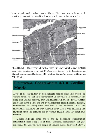
between  individual  cardiac  muscle  fibers.  The  clear  spaces  between  the

               myofibrils represent the branching features of different cardiac muscle fibers.


               FIGURE 8.13 Ultrastructure of cardiac muscle in longitudinal section. ×24,800.

               Used  with  permission  from  Cui  D.  Atlas  of  Histology  with  Functional  and
               Clinical Correlations. Baltimore, MD: Wolters Kluwer/Lippincott Williams and
               Wilkins; 2011.


                  FUNCTIONAL  CORRELATIONS  8.3  ■  Cardiac


                  Muscle


                 Although the organization of the contractile proteins (actin and myosin) in
                 cardiac  myofibers  and  their  arrangement  in  sarcomeres  is  essentially  the
                 same as in skeletal muscles, there are important differences. The T tubules
                 are located at the Z lines and are much larger than those in skeletal muscles.

                 Furthermore,  the  sarcoplasmic  reticulum  is  less  developed.  Also,  the
                 mitochondria are larger and more abundant in the cardiac cells indicating the
                 increased  metabolic  demands  on  the  cardiac  muscle  fibers  for  continuous

                 function.

                     Cardiac  cells  are  joined  end  to  end  by  specialized,  interdigitating
                 intercalated  discs  composed  of  fascia  adherens,  desmosomes,  and  gap
                 junctions.  The  gap  junctions  couple  all  cardiac  muscle  fibers  and  allow  a


                                                          313