
Page 319 - Atlas of Histology with Functional Correlations
P. 319
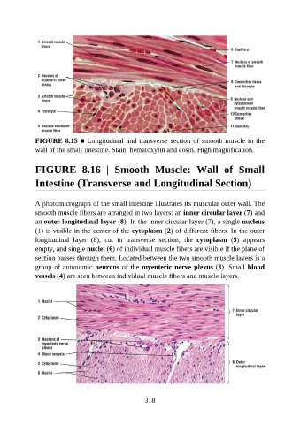
FIGURE 8.15 ■ Longitudinal and transverse section of smooth muscle in the
               wall of the small intestine. Stain: hematoxylin and eosin. High magnification.


               FIGURE  8.16  |  Smooth  Muscle:  Wall  of  Small

               Intestine (Transverse and Longitudinal Section)


               A photomicrograph of the small intestine illustrates its muscular outer wall. The
               smooth muscle fibers are arranged in two layers: an inner circular layer (7) and
               an outer longitudinal layer (8). In the inner circular layer (7), a single nucleus

               (1) is visible in the center of the cytoplasm (2) of different fibers. In the outer
               longitudinal  layer  (8),  cut  in  transverse  section,  the  cytoplasm  (5)  appears
               empty, and single nuclei (6) of individual muscle fibers are visible if the plane of
               section passes through them. Located between the two smooth muscle layers is a

               group of autonomic neurons of the myenteric nerve plexus (3). Small blood
               vessels (4) are seen between individual muscle fibers and muscle layers.


                                                          318