
Page 321 - Atlas of Histology with Functional Correlations
P. 321
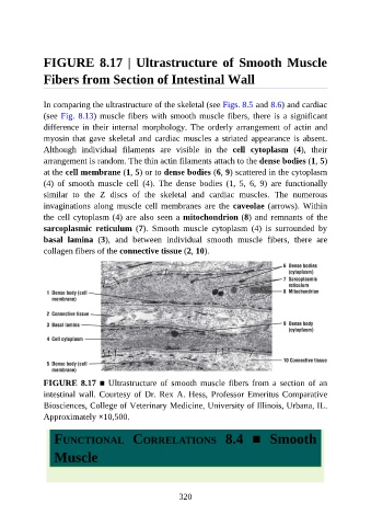
FIGURE  8.17  |  Ultrastructure  of  Smooth  Muscle

               Fibers from Section of Intestinal Wall


               In comparing the ultrastructure of the skeletal (see Figs. 8.5 and 8.6) and cardiac
               (see Fig. 8.13) muscle fibers with smooth muscle fibers, there is a significant

               difference  in  their  internal  morphology.  The  orderly  arrangement  of  actin  and
               myosin that gave skeletal and cardiac muscles a striated appearance is absent.
               Although  individual  filaments  are  visible  in  the  cell  cytoplasm  (4),  their

               arrangement is random. The thin actin filaments attach to the dense bodies (1, 5)
               at the cell membrane (1, 5) or to dense bodies (6, 9) scattered in the cytoplasm
               (4)  of  smooth  muscle  cell  (4).  The  dense  bodies  (1,  5,  6,  9)  are  functionally
               similar  to  the  Z  discs  of  the  skeletal  and  cardiac  muscles.  The  numerous
               invaginations along muscle cell membranes are the caveolae (arrows). Within

               the cell cytoplasm (4) are also seen a mitochondrion (8) and remnants of the
               sarcoplasmic  reticulum  (7).  Smooth  muscle  cytoplasm  (4)  is  surrounded  by
               basal  lamina  (3),  and  between  individual  smooth  muscle  fibers,  there  are

               collagen fibers of the connective tissue (2, 10).


               FIGURE 8.17 ■  Ultrastructure  of  smooth  muscle  fibers  from  a  section  of  an
               intestinal wall. Courtesy of Dr. Rex A. Hess, Professor Emeritus Comparative
               Biosciences, College of Veterinary Medicine, University of Illinois, Urbana, IL.

               Approximately ×10,500.


                  FUNCTIONAL  CORRELATIONS  8.4  ■  Smooth

                  Muscle


                                                          320