
Page 318 - Atlas of Histology with Functional Correlations
P. 318
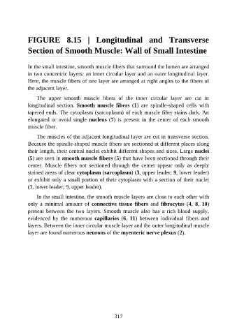
FIGURE  8.15  |  Longitudinal  and  Transverse

               Section of Smooth Muscle: Wall of Small Intestine


               In the small intestine, smooth muscle fibers that surround the lumen are arranged
               in two concentric layers: an inner circular layer and an outer longitudinal layer.

               Here, the muscle fibers of one layer are arranged at right angles to the fibers of
               the adjacent layer.

                   The  upper  smooth  muscle  fibers  of  the  inner  circular  layer  are  cut  in
               longitudinal  section.  Smooth  muscle  fibers  (1)  are  spindle-shaped  cells  with

               tapered ends. The cytoplasm (sarcoplasm) of each muscle fiber stains dark. An
               elongated or ovoid single nucleus (7) is present in the center of each smooth
               muscle fiber.

                   The muscles of the adjacent longitudinal layer are cut in transverse section.
               Because the spindle-shaped muscle fibers are sectioned at different places along

               their length, their central nuclei exhibit different shapes and sizes. Large nuclei
               (5) are seen in smooth muscle fibers (5) that have been sectioned through their
               center.  Muscle  fibers  not  sectioned  through  the  center  appear  only  as  deeply

               stained areas of clear cytoplasm (sarcoplasm) (3, upper leader; 9, lower leader)
               or exhibit only a small portion of their cytoplasm with a section of their nuclei
               (3, lower leader; 9, upper leader).

                   In the small intestine, the smooth muscle layers are close to each other with
               only  a  minimal  amount  of  connective  tissue  fibers  and  fibrocytes  (4,  8,  10)

               present  between  the  two  layers.  Smooth  muscle  also  has  a  rich  blood  supply,
               evidenced  by  the  numerous  capillaries  (6,  11)  between  individual  fibers  and
               layers. Between the inner circular muscle layer and the outer longitudinal muscle
               layer are found numerous neurons of the myenteric nerve plexus (2).


                                                          317