
Page 376 - Atlas of Histology with Functional Correlations
P. 376
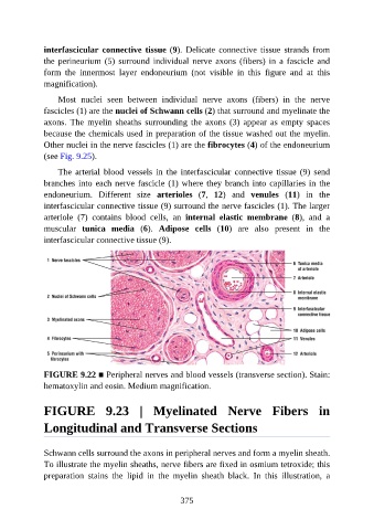
interfascicular  connective  tissue (9).  Delicate  connective  tissue  strands  from

               the  perineurium  (5)  surround  individual  nerve  axons  (fibers)  in  a  fascicle  and
               form  the  innermost  layer  endoneurium  (not  visible  in  this  figure  and  at  this
               magnification).

                   Most  nuclei  seen  between  individual  nerve  axons  (fibers)  in  the  nerve
               fascicles (1) are the nuclei of Schwann cells (2) that surround and myelinate the

               axons.  The  myelin  sheaths  surrounding  the  axons  (3)  appear  as  empty  spaces
               because the chemicals used in preparation of the tissue washed out the myelin.
               Other nuclei in the nerve fascicles (1) are the fibrocytes (4) of the endoneurium
               (see Fig. 9.25).


                   The  arterial  blood  vessels  in  the  interfascicular  connective  tissue  (9)  send
               branches into each nerve fascicle (1) where they branch into capillaries in the
               endoneurium.  Different  size  arterioles  (7,  12)  and  venules  (11)  in  the
               interfascicular connective tissue (9) surround the nerve fascicles (1). The larger
               arteriole  (7)  contains  blood  cells,  an  internal  elastic  membrane  (8),  and  a

               muscular  tunica  media  (6).  Adipose  cells  (10)  are  also  present  in  the
               interfascicular connective tissue (9).


               FIGURE 9.22 ■ Peripheral nerves and blood vessels (transverse section). Stain:
               hematoxylin and eosin. Medium magnification.


               FIGURE  9.23  |  Myelinated  Nerve  Fibers  in

               Longitudinal and Transverse Sections


               Schwann cells surround the axons in peripheral nerves and form a myelin sheath.

               To illustrate the myelin sheaths, nerve fibers are fixed in osmium tetroxide; this
               preparation  stains  the  lipid  in  the  myelin  sheath  black.  In  this  illustration,  a


                                                          375