
Page 379 - Atlas of Histology with Functional Correlations
P. 379
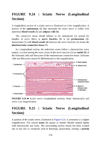
FIGURE  9.24  |  Sciatic  Nerve  (Longitudinal

               Section)


               A longitudinal section of a sciatic nerve is illustrated at a low magnification. A
               portion  of  the  epineurium  (1)  that  surrounds  the  entire  nerve  is  visible  with

               numerous blood vessels (5) and adipose cells (6).

                   The  connective  tissue  sheath  inferior  to  the  epineurium  (1)  around  the
               bundles  of  nerve  fibers  or  nerve  fascicles  (3)  is  the  perineurium  (2).
               Epineurium (1) with blood vessels (4) between the nerve fascicles (3) forms the
               interfascicular connective tissue (7).


                   In a longitudinal section, the individual axons follow a characteristic wavy
               pattern. Located among the wavy axons in the nerve fascicle (3) are nuclei (8) of
               the Schwann cells and fibrocytes of the endoneurium connective tissue. Schwann
               cells and fibrocytes cannot be differentiated at this magnification.


               FIGURE  9.24  ■  Sciatic  nerve  (longitudinal  section).  Stain:  hematoxylin  and
               eosin. Low magnification.


               FIGURE  9.25  |  Sciatic  Nerve  (Longitudinal

               Section)


               A portion of the sciatic nerve, illustrated in Figure 9.24, is presented at a higher

               magnification.  The  central  axons (1)  appear  as  slender  threads  stained  lightly
               with hematoxylin  and  eosin.  The  surrounding myelin  sheath  has  been  washed
               out  or  lost  due  to  chemicals  used  in  histologic  preparation,  leaving  a  protein


                                                          378