
Page 380 - Atlas of Histology with Functional Correlations
P. 380
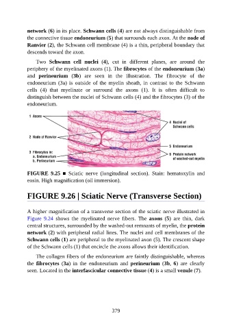
network (6) in its place. Schwann cells (4) are not always distinguishable from

               the connective tissue endoneurium (5) that surrounds each axon. At the node of
               Ranvier (2), the Schwann cell membrane (4) is a thin, peripheral boundary that
               descends toward the axon.

                   Two  Schwann  cell  nuclei  (4),  cut  in  different  planes,  are  around  the
               periphery of the myelinated axons (1). The fibrocytes of the endoneurium (3a)

               and  perineurium  (3b)  are  seen  in  the  illustration.  The  fibrocyte  of  the
               endoneurium  (3a)  is  outside  of  the  myelin  sheath,  in  contrast  to  the  Schwann
               cells  (4)  that  myelinate  or  surround  the  axons  (1).  It  is  often  difficult  to
               distinguish between the nuclei of Schwann cells (4) and the fibrocytes (3) of the

               endoneurium.


               FIGURE  9.25  ■  Sciatic  nerve  (longitudinal  section).  Stain:  hematoxylin  and
               eosin. High magnification (oil immersion).


               FIGURE 9.26 | Sciatic Nerve (Transverse Section)


               A higher magnification of a transverse section of the sciatic nerve illustrated in
               Figure  9.24  shows  the  myelinated  nerve  fibers.  The  axons  (5)  are  thin,  dark

               central structures, surrounded by the washed-out remnants of myelin, the protein
               network (2) with peripheral radial lines. The nuclei and cell membranes of the
               Schwann cells (1) are peripheral to the myelinated axon (5). The crescent shape
               of the Schwann cells (1) that encircle the axons allows their identification.

                   The collagen fibers of the endoneurium are faintly distinguishable, whereas

               the  fibrocytes  (3a)  in  the  endoneurium  and  perineurium  (3b,  6)  are  clearly
               seen. Located in the interfascicular connective tissue (4) is a small venule (7).


                                                          379