
Page 385 - Atlas of Histology with Functional Correlations
P. 385
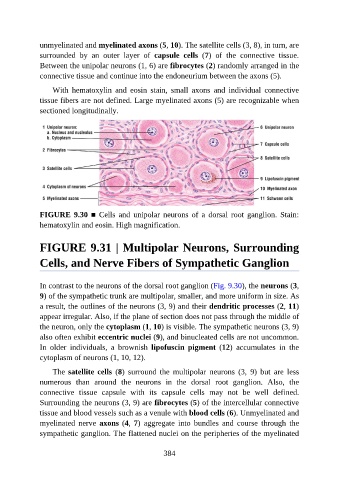
unmyelinated and myelinated axons (5, 10). The satellite cells (3, 8), in turn, are

               surrounded  by  an  outer  layer  of  capsule  cells  (7)  of  the  connective  tissue.
               Between the unipolar neurons (1, 6) are fibrocytes (2) randomly arranged in the
               connective tissue and continue into the endoneurium between the axons (5).

                   With  hematoxylin  and  eosin  stain,  small  axons  and  individual  connective
               tissue fibers are not defined. Large myelinated axons (5) are recognizable when

               sectioned longitudinally.


               FIGURE 9.30 ■  Cells  and  unipolar  neurons  of  a  dorsal  root  ganglion.  Stain:

               hematoxylin and eosin. High magnification.


               FIGURE 9.31 | Multipolar Neurons, Surrounding


               Cells, and Nerve Fibers of Sympathetic Ganglion


               In contrast to the neurons of the dorsal root ganglion (Fig. 9.30), the neurons (3,
               9) of the sympathetic trunk are multipolar, smaller, and more uniform in size. As
               a result, the outlines of the neurons (3, 9) and their dendritic processes (2, 11)

               appear irregular. Also, if the plane of section does not pass through the middle of
               the neuron, only the cytoplasm (1, 10) is visible. The sympathetic neurons (3, 9)
               also often exhibit eccentric nuclei (9), and binucleated cells are not uncommon.

               In  older  individuals,  a  brownish  lipofuscin  pigment  (12)  accumulates  in  the
               cytoplasm of neurons (1, 10, 12).

                   The satellite cells  (8)  surround  the  multipolar  neurons  (3,  9)  but  are  less
               numerous  than  around  the  neurons  in  the  dorsal  root  ganglion.  Also,  the
               connective  tissue  capsule  with  its  capsule  cells  may  not  be  well  defined.

               Surrounding the neurons (3, 9) are fibrocytes (5) of the intercellular connective
               tissue and blood vessels such as a venule with blood cells (6). Unmyelinated and
               myelinated  nerve  axons (4, 7)  aggregate  into  bundles  and  course  through  the
               sympathetic ganglion. The flattened nuclei on the peripheries of the myelinated


                                                          384