
Page 383 - Atlas of Histology with Functional Correlations
P. 383
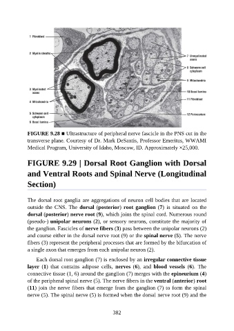
FIGURE 9.28 ■ Ultrastructure of peripheral nerve fascicle in the PNS cut in the
               transverse plane. Courtesy of Dr. Mark DeSantis, Professor Emeritus, WWAMI
               Medical Program, University of Idaho, Moscow, ID. Approximately ×25,000.


               FIGURE 9.29 | Dorsal Root Ganglion with Dorsal

               and Ventral Roots and Spinal Nerve (Longitudinal


               Section)


               The dorsal root ganglia are aggregations of neuron cell bodies that are located
               outside  the  CNS.  The  dorsal (posterior) root ganglion (7)  is  situated  on  the

               dorsal (posterior) nerve root (9), which joins the spinal cord. Numerous round
               (pseudo-) unipolar neurons (2), or sensory neurons, constitute the majority of
               the ganglion. Fascicles of nerve fibers (3) pass between the unipolar neurons (2)
               and course either in the dorsal nerve root (9) or the spinal nerve (5). The nerve
               fibers (3) represent the peripheral processes that are formed by the bifurcation of

               a single axon that emerges from each unipolar neuron (2).

                   Each dorsal root ganglion (7) is enclosed by an irregular connective tissue
               layer  (1)  that  contains  adipose  cells,  nerves  (6),  and  blood  vessels  (6).  The
               connective tissue (1, 6) around the ganglion (7) merges with the epineurium (4)

               of the peripheral spinal nerve (5). The nerve fibers in the ventral (anterior) root
               (11) join the nerve fibers that emerge from the ganglion (7) to form the spinal
               nerve (5). The spinal nerve (5) is formed when the dorsal nerve root (9) and the


                                                          382