
Page 386 - Atlas of Histology with Functional Correlations
P. 386
axons (4, 7) are the Schwann cells (4, 7). These nerve fibers represent the
preganglionic axons, postganglionic visceral efferent axons, and visceral afferent
axons.
FIGURE 9.31 ■ Multipolar neurons, surrounding cells, and nerve fibers of a
sympathetic ganglion. Stain: hematoxylin and eosin. High magnification.
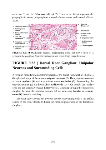
FIGURE 9.32 | Dorsal Root Ganglion: Unipolar
Neurons and Surrounding Cells
A medium-magnification photomicrograph of the dorsal root ganglion illustrates
the spherical shape of the sensory unipolar neurons (2). The cytoplasm contains
a central nucleus (6) and a prominent dense nucleolus (5). Surrounding the
unipolar neurons (2) are the smaller satellite cells (1). Cells outside the satellite
cells are the connective tissue fibrocytes (3). Coursing through the dorsal root
ganglion between the unipolar neurons (2) are numerous bundles of sensory
axons (4) from the periphery.
The clear space around the neurons and the surrounding cells is an artifact
caused by the tissue shrinkage during the chemical preparation of the dorsal root
ganglion.
385