
Page 381 - Atlas of Histology with Functional Correlations
P. 381
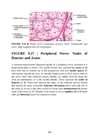
FIGURE  9.26  ■  Sciatic  nerve  (transverse  section).  Stain:  hematoxylin  and

               eosin. High magnification (oil immersion).


               FIGURE  9.27  |  Peripheral  Nerve:  Nodes  of


               Ranvier and Axons


               A medium-magnification photomicrograph of a peripheral nerve sectioned in a
               longitudinal plane is shown. The myelin sheaths that surround the axons (2, 8)
               have been lost or washed out in this preparation, and only myelin spaces (7)

               with protein network are seen. A centrally located axon (2, 8) is seen in some of
               the  nerve  fibers  that  exhibited  myelin  sheaths.  At  regular  intervals  along  the
               axon  are  indentations  or  in  the  myelin  sheaths.  These  represent  the  nodes  of
               Ranvier (1, 9). These sites indicate the edges of two different myelin sheaths

               that enclose the axon. A possible Schwann cell nucleus (3) is seen with one of
               the axons (2, 8) and a thin, blue connective tissue layer endoneurium (6) around
               some of the axons (2, 8). Outside of the axons (2, 8) are capillary (4) with blood
               cells and fibrocytes (5) of the connective tissue.


                                                          380