
Page 382 - Atlas of Histology with Functional Correlations
P. 382
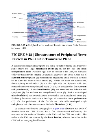
FIGURE 9.27 ■ Peripheral nerve: nodes of Ranvier and axons. Stain: Masson
               trichrome. ×100.


               FIGURE 9.28 | Ultrastructure of Peripheral Nerve


               Fascicle in PNS Cut in Transverse Plane


               A transmission electron micrograph of a nerve fascicle sectioned in a transverse
               plane  shows  two  large  myelinated  axons  (3)  on  the  left  side  and  small

               unmyelinated axons (7) on the right side. In contrast to the CNS, the Schwann
               cells only form myelin sheaths (2) around a section of one axon. A thin rim of
               Schwann cell cytoplasm (5) surrounds the myelinated axon, which is invested
               by  an  outer  thin  layer  of  basal  lamina  (6).  Within  the  axons  are  oval-shaped,

               dense-staining  mitochondria  (4).  On  the  right  side  are  Schwann  cells  that
               surround numerous unmyelinated axons (7) that are embedded in the Schwann
               cell cytoplasm (8). A thin basal lamina (10) also surrounds the Schwann cell
               cytoplasm  (8)  that  encloses  the  unmyelinated  axons  (7).  Similar  oval-shaped

               mitochondria (9) and neurofilaments are found in the unmyelinated axons (7).
               Enclosing  the  nerve  fascicle  is  a  thin  layer  of  connective  tissue  perineurium
               (12).  On  the  peripheries  of  the  fascicle  are  cells  with  developed  rough
               endoplasmic reticulum that are most likely the fibroblasts (1, 11).


                   A  transmission  electron  micrograph  of  Figure  9.19  illustrates  the  node  of
               Ranvier  from  the  CNS.  Except  for  a  few  ultrastructural  differences,  the
               structures  of  the  nodes  of  Ranvier  in  the  PNS  and  the  CNS  are  similar.  The
               nodes in the PNS are covered by the basal lamina, whereas the nodes in the
               CNS lack an overlying basal lamina.


                                                          381