
Page 377 - Atlas of Histology with Functional Correlations
P. 377
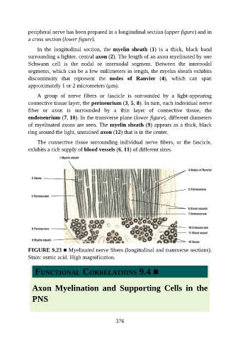
peripheral nerve has been prepared in a longitudinal section (upper figure) and in

               a cross section (lower figure).

                   In  the  longitudinal  section,  the  myelin  sheath  (1)  is  a  thick,  black  band
               surrounding a lighter, central axon (2). The length of an axon myelinated by one
               Schwann  cell  is  the  nodal  or  internodal  segment.  Between  the  internodal
               segments, which can be a few millimeters in length, the myelin sheath exhibits

               discontinuity  that  represent  the  nodes  of  Ranvier  (4),  which  can  span
               approximately 1 or 2 micrometers (μm).

                   A  group  of  nerve  fibers  or  fascicle  is  surrounded  by  a  light-appearing
               connective tissue layer, the perineurium (3, 5, 8). In turn, each individual nerve

               fiber  or  axon  is  surrounded  by  a  thin  layer  of  connective  tissue,  the
               endoneurium (7, 10). In the transverse plane (lower figure), different diameters
               of myelinated axons are seen. The myelin sheath (9) appears as a thick, black
               ring around the light, unstained axon (12) that is in the center.

                   The  connective  tissue  surrounding  individual  nerve  fibers,  or  the  fascicle,

               exhibits a rich supply of blood vessels (6, 11) of different sizes.


               FIGURE 9.23 ■ Myelinated nerve fibers (longitudinal and transverse sections).
               Stain: osmic acid. High magnification.


                  FUNCTIONAL CORRELATIONS 9.4 ■


                 Axon  Myelination  and  Supporting  Cells  in  the


                 PNS


                                                          376