
Page 384 - Atlas of Histology with Functional Correlations
P. 384
ventral (anterior) root (11) unite.
On emerging from the spinal cord, the dorsal (9) and ventral roots (11) are
surrounded by pia mater and an arachnoid sheath (8, 10). These become
continuous with the epineurium (4) of the spinal nerve (5). The perineurium
around the nerve fascicles (3) and the endoneurium around individual nerve
fibers in the spinal nerve (5) or in the ganglion (7) are not distinguishable at this
magnification.
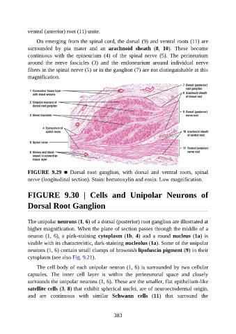
FIGURE 9.29 ■ Dorsal root ganglion, with dorsal and ventral roots, spinal
nerve (longitudinal section). Stain: hematoxylin and eosin. Low magnification.
FIGURE 9.30 | Cells and Unipolar Neurons of
Dorsal Root Ganglion
The unipolar neurons (1, 6) of a dorsal (posterior) root ganglion are illustrated at
higher magnification. When the plane of section passes through the middle of a
neuron (1, 6), a pink-staining cytoplasm (1b, 4) and a round nucleus (1a) is
visible with its characteristic, dark-staining nucleolus (1a). Some of the unipolar
neurons (1, 6) contain small clumps of brownish lipofuscin pigment (9) in their
cytoplasm (see also Fig. 9.21).
The cell body of each unipolar neuron (1, 6) is surrounded by two cellular
capsules. The inner cell layer is within the perineuronal space and closely
surrounds the unipolar neurons (1, 6). These are the smaller, flat epithelium-like
satellite cells (3, 8) that exhibit spherical nuclei, are of neuroectodermal origin,
and are continuous with similar Schwann cells (11) that surround the
383