
Page 395 - Atlas of Histology with Functional Correlations
P. 395
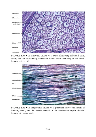
FIGURE  9.39  ■  A  transverse  section  of  a  nerve  illustrating  individual  cells,
               axons,  and  the  surrounding  connective  tissue.  Stain:  hematoxylin  and  eosin.
               Masson stain. ×100.


               FIGURE  9.40  ■  A  longitudinal  section  of  a  peripheral  nerve  with  nodes  of
               Ranvier,  axons,  and  the  protein  network  in  the  washed-out  myelin  sheaths.
               Masson trichrome. ×165.


                                                          394