
Page 400 - Atlas of Histology with Functional Correlations
P. 400
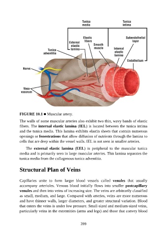
FIGURE 10.1 ■ Muscular artery.

               The walls of some muscular arteries also exhibit two thin, wavy bands of elastic
               fibers. The internal elastic lamina (IEL) is located between the tunica intima

               and the tunica media. This lamina exhibits elastin sheets that contain numerous
               openings or fenestrations that allow diffusion of nutrients through the lamina to
               cells that are deep within the vessel walls. IEL is not seen in smaller arteries.

                   The  external  elastic  lamina  (EEL)  is  peripheral  to  the  muscular  tunica

               media and is primarily seen in large muscular arteries. This lamina separates the
               tunica media from the collagenous tunica adventitia.


               Structural Plan of Veins


               Capillaries  unite  to  form  larger  blood  vessels  called  venules  that  usually
               accompany  arterioles.  Venous  blood  initially  flows  into  smaller  postcapillary
               venules and then into veins of increasing size. The veins are arbitrarily classified
               as small, medium, and large. Compared with arteries, veins are more numerous
               and have thinner walls, larger diameters, and greater structural variation. Blood

               that enters the veins is under low pressure. Small-sized and medium-sized veins,
               particularly veins in the extremities (arms and legs) and those that convey blood


                                                          399