
Page 405 - Atlas of Histology with Functional Correlations
P. 405
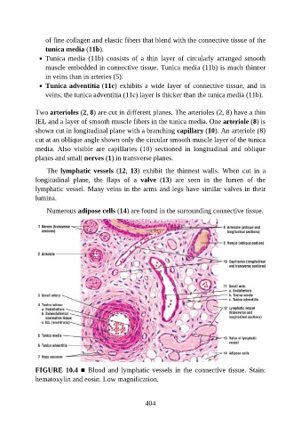
of fine collagen and elastic fibers that blend with the connective tissue of the

                   tunica media (11b).
                   Tunica  media  (11b)  consists  of  a  thin  layer  of  circularly  arranged  smooth
                   muscle embedded in connective tissue. Tunica media (11b) is much thinner
                   in veins than in arteries (5).
                   Tunica adventitia (11c) exhibits a wide layer of connective tissue, and in

                   veins, the tunica adventitia (11c) layer is thicker than the tunica media (11b).


               Two arterioles (2, 8) are cut in different planes. The arterioles (2, 8) have a thin
               IEL and a layer of smooth muscle fibers in the tunica media. One arteriole (8) is

               shown cut in longitudinal plane with a branching capillary (10). An arteriole (8)
               cut at an oblique angle shown only the circular smooth muscle layer of the tunica
               media.  Also  visible  are  capillaries  (10)  sectioned  in  longitudinal  and  oblique
               planes and small nerves (1) in transverse planes.

                   The lymphatic vessels (12, 13)  exhibit  the  thinnest  walls.  When  cut  in  a

               longitudinal  plane,  the  flaps  of  a  valve  (13)  are  seen  in  the  lumen  of  the
               lymphatic vessel. Many veins in the arms and legs have similar valves in their
               lumina.

                   Numerous adipose cells (14) are found in the surrounding connective tissue.


               FIGURE 10.4 ■  Blood  and  lymphatic  vessels  in  the  connective  tissue.  Stain:

               hematoxylin and eosin. Low magnification.


                                                          404