
Page 410 - Atlas of Histology with Functional Correlations
P. 410
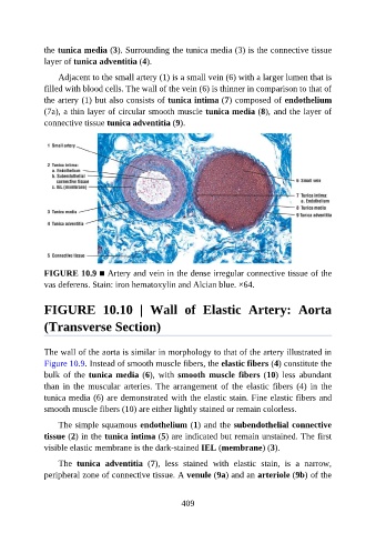
the tunica media (3). Surrounding the tunica media (3) is the connective tissue

               layer of tunica adventitia (4).

                   Adjacent to the small artery (1) is a small vein (6) with a larger lumen that is
               filled with blood cells. The wall of the vein (6) is thinner in comparison to that of
               the artery (1) but also consists of tunica intima (7) composed of endothelium
               (7a), a thin layer of circular smooth muscle tunica media (8), and the layer of

               connective tissue tunica adventitia (9).


               FIGURE 10.9 ■ Artery and vein in the dense irregular connective tissue of the

               vas deferens. Stain: iron hematoxylin and Alcian blue. ×64.


               FIGURE  10.10  |  Wall  of  Elastic  Artery:  Aorta


               (Transverse Section)


               The wall of the aorta is similar in morphology to that of the artery illustrated in
               Figure 10.9. Instead of smooth muscle fibers, the elastic fibers (4) constitute the
               bulk  of  the  tunica media (6),  with  smooth  muscle  fibers (10)  less  abundant

               than  in  the  muscular  arteries.  The  arrangement  of  the  elastic  fibers  (4)  in  the
               tunica media (6) are demonstrated with the elastic stain. Fine elastic fibers and
               smooth muscle fibers (10) are either lightly stained or remain colorless.

                   The simple squamous endothelium (1) and the subendothelial connective

               tissue (2) in the tunica intima (5) are indicated but remain unstained. The first
               visible elastic membrane is the dark-stained IEL (membrane) (3).

                   The  tunica  adventitia  (7),  less  stained  with  elastic  stain,  is  a  narrow,
               peripheral zone of connective tissue. A venule (9a) and an arteriole (9b) of the


                                                          409