
Page 412 - Atlas of Histology with Functional Correlations
P. 412
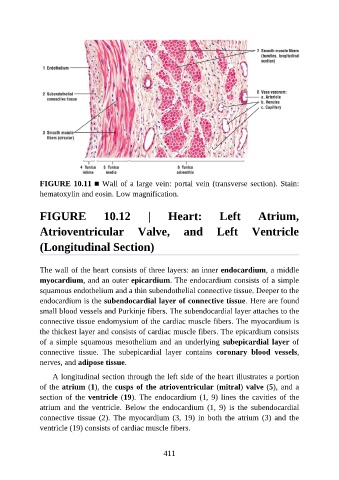
FIGURE 10.11 ■ Wall of a large vein: portal vein (transverse section). Stain:
               hematoxylin and eosin. Low magnification.


               FIGURE                10.12           |      Heart:            Left          Atrium,


               Atrioventricular  Valve,  and  Left  Ventricle

               (Longitudinal Section)


               The wall of the heart consists of three layers: an inner endocardium, a middle

               myocardium, and an outer epicardium. The endocardium consists of a simple
               squamous endothelium and a thin subendothelial connective tissue. Deeper to the
               endocardium is the subendocardial layer of connective tissue. Here are found

               small blood vessels and Purkinje fibers. The subendocardial layer attaches to the
               connective tissue endomysium of the cardiac muscle fibers. The myocardium is
               the thickest layer and consists of cardiac muscle fibers. The epicardium consists
               of a simple squamous mesothelium and an underlying subepicardial layer of
               connective  tissue.  The  subepicardial  layer  contains  coronary  blood  vessels,

               nerves, and adipose tissue.

                   A longitudinal section through the left side of the heart illustrates a portion
               of the atrium (1), the cusps of the atrioventricular (mitral) valve (5), and a
               section of the ventricle (19). The endocardium (1, 9) lines the cavities of the

               atrium  and  the  ventricle.  Below  the  endocardium  (1,  9)  is  the  subendocardial
               connective  tissue  (2).  The  myocardium  (3,  19)  in  both  the  atrium  (3)  and  the
               ventricle (19) consists of cardiac muscle fibers.


                                                          411