
Page 416 - Atlas of Histology with Functional Correlations
P. 416
(9) that surrounds the Purkinje fibers (6, 10). The characteristic features of
Purkinje fibers (6, 10) are visible in both longitudinal and transverse planes. In
transverse plane (6), the Purkinje fibers exhibit fewer peripheral myofibrils
leaving a perinuclear zone of comparatively clear sarcoplasm. A central nucleus
is seen in some transverse sections; in others, a central area of clear sarcoplasm
is seen, with the plane of section bypassing the nucleus.
The Purkinje fibers (6, 10) are located inferior to the endocardium (7),
which represents the endothelium of the heart cavities. The Purkinje fibers (6,
10) are different from typical cardiac muscle fibers (1, 3) in that the Purkinje
fibers (6, 10) are larger and show less intense staining.
The cardiac muscle fibers (1, 3) are connected to each other via the
prominent intercalated discs (4). The intercalated discs (4) are not observed in
the Purkinje fibers (6, 10). Instead, the Purkinje fibers (6, 10) are connected to
each other via desmosomes and gap junctions and eventually merge with cardiac
muscle fibers (1, 3).
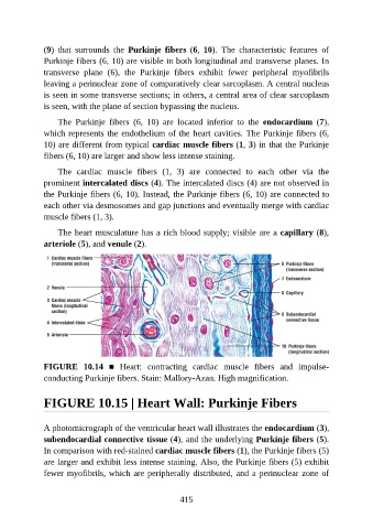
The heart musculature has a rich blood supply; visible are a capillary (8),
arteriole (5), and venule (2).
FIGURE 10.14 ■ Heart: contracting cardiac muscle fibers and impulse-
conducting Purkinje fibers. Stain: Mallory-Azan. High magnification.
FIGURE 10.15 | Heart Wall: Purkinje Fibers
A photomicrograph of the ventricular heart wall illustrates the endocardium (3),
subendocardial connective tissue (4), and the underlying Purkinje fibers (5).
In comparison with red-stained cardiac muscle fibers (1), the Purkinje fibers (5)
are larger and exhibit less intense staining. Also, the Purkinje fibers (5) exhibit
fewer myofibrils, which are peripherally distributed, and a perinuclear zone of
415