
Page 417 - Atlas of Histology with Functional Correlations
P. 417
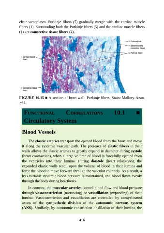
clear  sarcoplasm.  Purkinje  fibers  (5)  gradually  merge  with  the  cardiac  muscle

               fibers (1). Surrounding both the Purkinje fibers (5) and the cardiac muscle fibers
               (1) are connective tissue fibers (2).


               FIGURE 10.15 ■ A section of heart wall: Purkinje fibers. Stain: Mallory-Azan.
               ×64.


                  FUNCTIONAL                     CORRELATIONS                       10.1            ■


                  Circulatory System


                 Blood Vessels


                     The elastic arteries transport the ejected blood from the heart and move
                 it along the systemic vascular path. The presence of elastic fibers in their

                 walls allows the elastic arteries to greatly expand in diameter during systole
                 (heart contraction), when a large volume of blood is forcefully ejected from
                 the  ventricles  into  their  lumina.  During  diastole  (heart  relaxation),  the
                 expanded elastic walls recoil upon the volume of blood in their lumina and
                 force the blood to move forward through the vascular channels. As a result, a

                 less variable systemic blood pressure is maintained, and blood flows evenly
                 through the body during heartbeats.

                     In contrast, the muscular arteries control blood flow and blood pressure
                 through vasoconstriction (narrowing) or vasodilation  (expanding)  of  their

                 lumina.  Vasoconstriction  and  vasodilation  are  controlled  by  unmyelinated
                 axons  of  the  sympathetic  division  of  the  autonomic  nervous  system
                 (ANS). Similarly, by autonomic constriction or dilation of their lumina, the


                                                          416