
Page 414 - Atlas of Histology with Functional Correlations
P. 414
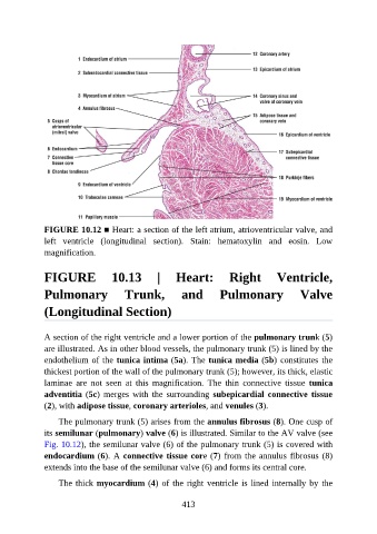
FIGURE 10.12 ■ Heart: a section of the left atrium, atrioventricular valve, and
               left  ventricle  (longitudinal  section).  Stain:  hematoxylin  and  eosin.  Low
               magnification.


               FIGURE  10.13  |  Heart:  Right  Ventricle,

               Pulmonary                Trunk,            and         Pulmonary                 Valve

               (Longitudinal Section)


               A section of the right ventricle and a lower portion of the pulmonary trunk (5)
               are illustrated. As in other blood vessels, the pulmonary trunk (5) is lined by the
               endothelium of the tunica intima (5a). The tunica media (5b) constitutes the
               thickest portion of the wall of the pulmonary trunk (5); however, its thick, elastic

               laminae  are  not  seen  at  this  magnification.  The  thin  connective  tissue  tunica
               adventitia (5c)  merges  with  the  surrounding  subepicardial  connective  tissue
               (2), with adipose tissue, coronary arterioles, and venules (3).

                   The pulmonary trunk (5) arises from the annulus fibrosus (8). One cusp of

               its semilunar (pulmonary) valve (6) is illustrated. Similar to the AV valve (see
               Fig. 10.12), the semilunar valve (6) of the pulmonary trunk (5) is covered with
               endocardium (6). A connective tissue core (7) from the annulus fibrosus (8)
               extends into the base of the semilunar valve (6) and forms its central core.

                   The thick myocardium (4) of the right ventricle is lined internally by the


                                                          413