
Page 409 - Atlas of Histology with Functional Correlations
P. 409
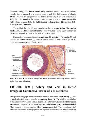
muscular  artery,  the  tunica  media  (1b),  contains  several  layers  of  smooth

               muscle  fibers,  arranged  in  a  circular  pattern,  and  thin  dark  strands  of  elastic
               fibers (1b). On the periphery of the tunica media (1b) is the less conspicuous
               EEL  (1c).  Surrounding  the  artery  is  the  connective  tissue  tunica  adventitia
               (1d),  which  contains  both  the  light-staining  collagen fibers (2)  and  the  dark-
               staining elastic fibers (3).


                   The wall of the vein (4) also contains the layers tunica intima (4a), tunica
               media (4b), and tunica adventitia (4c). However, these three layers in the vein
               (4) are not as thick as those in the wall of the artery (1).

                   Surrounding both vessels are the capillary (5), arteriole (7), venule (6), and

               cells of the adipose tissue (8). Present in the lumina of both vessels (1, 4) are
               numerous erythrocytes and leukocytes.


               FIGURE  10.8  ■  Muscular  artery  and  vein  (transverse  section).  Stain:  elastic
               stain. Low magnification.


               FIGURE  10.9  |  Artery  and  Vein  in  Dense

               Irregular Connective Tissue of Vas Deferens


               This photomicrograph illustrates the differences between a small artery (1) and
               a small vein (6) in dense irregular connective tissue (5). The small artery (1) has
               a thick muscular wall and a small lumen. The arterial wall consists of the tunica
               intima (2), composed of an inner layer of endothelium (2a), a subendothelial

               (2b)  connective  tissue,  and  an  IEL  (membrane)  (2c).  This  membrane  (2c)
               separates the tunica intima (2) from the layer of circular smooth muscle fibers of


                                                          408