
Page 406 - Atlas of Histology with Functional Correlations
P. 406
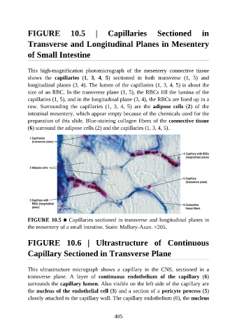
FIGURE  10.5  |  Capillaries  Sectioned  in

               Transverse and Longitudinal Planes in Mesentery

               of Small Intestine


               This  high-magnification  photomicrograph  of  the  mesentery  connective  tissue
               shows  the  capillaries  (1,  3,  4,  5)  sectioned  in  both  transverse  (1,  5)  and
               longitudinal planes (3, 4). The lumen of the capillaries (1, 3, 4, 5) is about the
               size of an RBC. In the transverse plane (1, 5), the RBCs fill the lumina of the
               capillaries (1, 5), and in the longitudinal plane (3, 4), the RBCs are lined up in a

               row.  Surrounding  the  capillaries  (1,  3,  4,  5)  are  the  adipose  cells  (2)  of  the
               intestinal mesentery, which appear empty because of the chemicals used for the
               preparation of this slide. Blue-staining collagen fibers of the connective tissue

               (6) surround the adipose cells (2) and the capillaries (1, 3, 4, 5).


               FIGURE 10.5 ■ Capillaries sectioned in transverse and longitudinal planes in

               the mesentery of a small intestine. Stain: Mallory-Azan. ×205.


               FIGURE  10.6  |  Ultrastructure  of  Continuous


               Capillary Sectioned in Transverse Plane


               This  ultrastructure  micrograph  shows  a  capillary  in  the  CNS,  sectioned  in  a
               transverse  plane.  A  layer  of  continuous  endothelium  of  the  capillary  (6)
               surrounds the capillary lumen. Also visible on the left side of the capillary are

               the nucleus of the endothelial cell (3) and a section of a pericyte process (5)
               closely attached to the capillary wall. The capillary endothelium (6), the nucleus


                                                          405