Page 407 - Atlas of Histology with Functional Correlations
P. 407
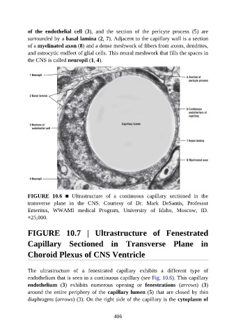
of  the  endothelial  cell  (3),  and  the  section  of  the  pericyte  process  (5)  are

               surrounded by a basal lamina (2, 7). Adjacent to the capillary wall is a section
               of a myelinated axon (8) and a dense meshwork of fibers from axons, dendrites,
               and astrocytic endfeet of glial cells. This neural meshwork that fills the spaces in
               the CNS is called neuropil (1, 4).


               FIGURE  10.6  ■  Ultrastructure  of  a  continuous  capillary  sectioned  in  the
               transverse  plane  in  the  CNS.  Courtesy  of  Dr.  Mark  DeSantis,  Professor
               Emeritus,  WWAMI  medical  Program,  University  of  Idaho,  Moscow,  ID.

               ×25,000.


               FIGURE  10.7  |  Ultrastructure  of  Fenestrated


               Capillary  Sectioned  in  Transverse  Plane  in

               Choroid Plexus of CNS Ventricle


               The  ultrastructure  of  a  fenestrated  capillary  exhibits  a  different  type  of

               endothelium that is seen in a continuous capillary (see Fig. 10.6). This capillary
               endothelium  (3)  exhibits  numerous  opening  or  fenestrations  (arrows)  (3)
               around the entire periphery of the capillary lumen (5) that are closed by thin
               diaphragms (arrows) (3). On the right side of the capillary is the cytoplasm of


                                                          406

