
Page 408 - Atlas of Histology with Functional Correlations
P. 408
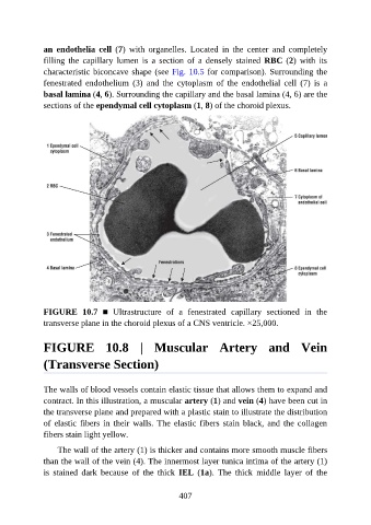
an  endothelia  cell  (7)  with  organelles.  Located  in  the  center  and  completely

               filling  the  capillary  lumen  is  a  section  of  a  densely  stained  RBC (2)  with  its
               characteristic biconcave shape (see Fig. 10.5 for comparison). Surrounding the
               fenestrated  endothelium  (3)  and  the  cytoplasm  of  the  endothelial  cell  (7)  is  a
               basal lamina (4, 6). Surrounding the capillary and the basal lamina (4, 6) are the
               sections of the ependymal cell cytoplasm (1, 8) of the choroid plexus.


               FIGURE  10.7  ■  Ultrastructure  of  a  fenestrated  capillary  sectioned  in  the
               transverse plane in the choroid plexus of a CNS ventricle. ×25,000.


               FIGURE  10.8  |  Muscular  Artery  and  Vein

               (Transverse Section)


               The walls of blood vessels contain elastic tissue that allows them to expand and

               contract. In this illustration, a muscular artery (1) and vein (4) have been cut in
               the transverse plane and prepared with a plastic stain to illustrate the distribution
               of elastic fibers in their walls. The elastic fibers stain black, and the collagen
               fibers stain light yellow.


                   The wall of the artery (1) is thicker and contains more smooth muscle fibers
               than the wall of the vein (4). The innermost layer tunica intima of the artery (1)
               is  stained  dark  because  of  the  thick  IEL  (1a).  The  thick  middle  layer  of  the


                                                          407