
Page 570 - Atlas of Histology with Functional Correlations
P. 570
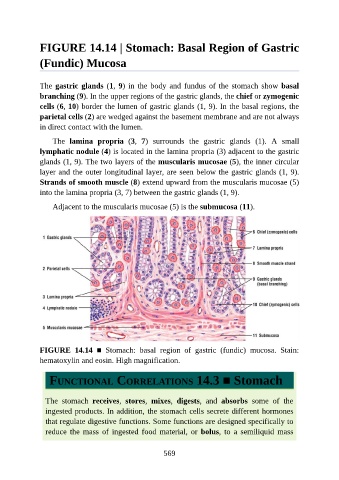
FIGURE 14.14 | Stomach: Basal Region of Gastric

               (Fundic) Mucosa


               The gastric glands (1, 9) in the body and fundus of the stomach show basal
               branching (9). In the upper regions of the gastric glands, the chief or zymogenic

               cells (6, 10) border the lumen of gastric glands (1, 9). In the basal regions, the
               parietal cells (2) are wedged against the basement membrane and are not always
               in direct contact with the lumen.

                   The  lamina  propria  (3,  7)  surrounds  the  gastric  glands  (1).  A  small
               lymphatic nodule (4) is located in the lamina propria (3) adjacent to the gastric

               glands (1, 9). The two layers of the muscularis mucosae (5), the inner circular
               layer and the outer longitudinal layer, are seen below the gastric glands (1, 9).
               Strands of smooth muscle (8) extend upward from the muscularis mucosae (5)

               into the lamina propria (3, 7) between the gastric glands (1, 9).

                   Adjacent to the muscularis mucosae (5) is the submucosa (11).


               FIGURE  14.14  ■  Stomach:  basal  region  of  gastric  (fundic)  mucosa.  Stain:
               hematoxylin and eosin. High magnification.


                  FUNCTIONAL CORRELATIONS 14.3 ■ Stomach


                 The  stomach  receives,  stores,  mixes,  digests,  and  absorbs  some  of  the

                 ingested products. In addition, the stomach cells secrete different hormones
                 that regulate digestive functions. Some functions are designed specifically to
                 reduce  the  mass  of  ingested  food  material,  or  bolus,  to  a  semiliquid  mass


                                                          569