
Page 568 - Atlas of Histology with Functional Correlations
P. 568
esophageal–stomach junction are the cardiac glands. The pylorus is the most
inferior, funnel-shaped region of the stomach that terminates at the border of
the small intestine called the duodenum. In the cardiac, the gastric pits are
shallow, whereas in the pylorus, the gastric pits are deep. However, the
histology of gastric glands in both regions is similar, and the cells are
predominantly mucus secreting.
In contrast, the gastric glands in the fundus and body exhibit different
histology and contain three major cell types. Located in gastric glands near
the gastric pits are the mucous neck cells. The large polygonal cells with
eosinophilic cytoplasm are the parietal cells that are primarily located in the
upper half of the gastric glands, squeezed between gland cells. Located
predominantly in the lower region of the gastric glands are basophilic
staining cuboidal chief (zymogenic) cells.
In addition to cells in gastric glands, the mucosa of the digestive tract
also contains enteroendocrine or gastrointestinal endocrine cells that are
part of the diffuse neuroendocrine system. These cells are distributed in
different digestive organs and are located among and between exocrine cells.
Unless digestive organs are prepared with special stains, the diffuse
neuroendocrine cells are difficult to see in normal histologic sections.
In addition, there are undifferentiated stem cells in the neck regions of
the gastric glands that continuously renew the cells in the gastric mucosa.
These stem cells move upward to replace lost or worn-out surface cells or
downward to replace the cells deep in the glands.
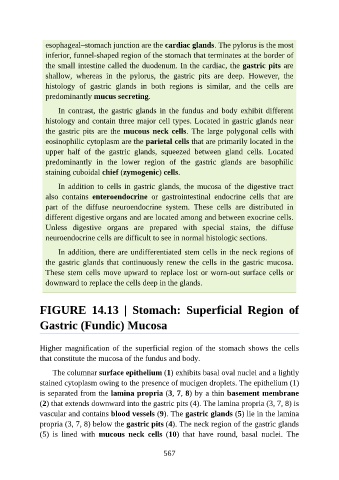
FIGURE 14.13 | Stomach: Superficial Region of
Gastric (Fundic) Mucosa
Higher magnification of the superficial region of the stomach shows the cells
that constitute the mucosa of the fundus and body.
The columnar surface epithelium (1) exhibits basal oval nuclei and a lightly
stained cytoplasm owing to the presence of mucigen droplets. The epithelium (1)
is separated from the lamina propria (3, 7, 8) by a thin basement membrane
(2) that extends downward into the gastric pits (4). The lamina propria (3, 7, 8) is
vascular and contains blood vessels (9). The gastric glands (5) lie in the lamina
propria (3, 7, 8) below the gastric pits (4). The neck region of the gastric glands
(5) is lined with mucous neck cells (10) that have round, basal nuclei. The
567