
Page 567 - Atlas of Histology with Functional Correlations
P. 567
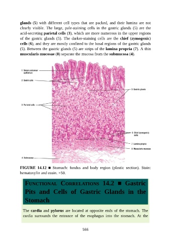
glands (5)  with  different  cell  types  that  are  packed,  and  their  lumina  are  not

               clearly  visible.  The  large,  pale-staining  cells  in  the  gastric  glands  (5)  are  the
               acid-secreting parietal cells (3), which are more numerous in the upper regions
               of  the  gastric  glands  (5).  The  darker-staining  cells  are  the  chief  (zymogenic)
               cells (6), and they are mostly confined to the basal regions of the gastric glands
               (5). Between the gastric glands (5) are strips of the lamina propria (7). A thin

               muscularis mucosae (8) separate the mucosa from the submucosa (4).


               FIGURE  14.12  ■  Stomach:  fundus  and  body  region  (plastic  section).  Stain:
               hematoxylin and eosin. ×50.


                  FUNCTIONAL  CORRELATIONS  14.2  ■  Gastric


                  Pits  and  Cells  of  Gastric  Glands  in  the


                  Stomach


                 The cardia  and  pylorus  are  located  at  opposite  ends  of  the  stomach.  The

                 cardia  surrounds  the  entrance  of  the  esophagus  into  the  stomach.  At  the


                                                          566