
Page 569 - Atlas of Histology with Functional Correlations
P. 569
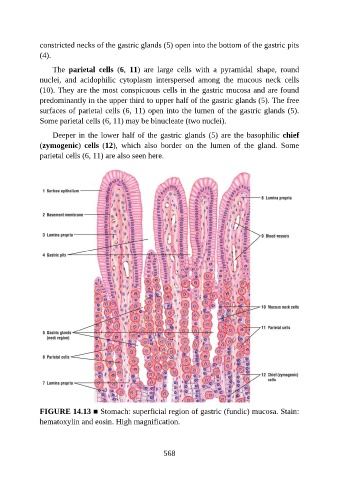
constricted necks of the gastric glands (5) open into the bottom of the gastric pits

               (4).

                   The  parietal  cells  (6,  11)  are  large  cells  with  a  pyramidal  shape,  round
               nuclei,  and  acidophilic  cytoplasm  interspersed  among  the  mucous  neck  cells
               (10). They are the most conspicuous cells in the gastric mucosa and are found
               predominantly in the upper third to upper half of the gastric glands (5). The free

               surfaces of parietal cells (6, 11) open into the lumen of the gastric glands (5).
               Some parietal cells (6, 11) may be binucleate (two nuclei).

                   Deeper in the lower half of the gastric glands (5) are the basophilic chief
               (zymogenic)  cells  (12),  which  also  border  on  the  lumen  of  the  gland.  Some

               parietal cells (6, 11) are also seen here.


               FIGURE 14.13 ■ Stomach: superficial region of gastric (fundic) mucosa. Stain:
               hematoxylin and eosin. High magnification.


                                                          568