Page 565 - Atlas of Histology with Functional Correlations
P. 565
hematoxylin and eosin. Low magnification.


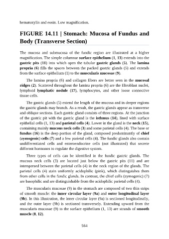
               FIGURE 14.11 | Stomach: Mucosa of Fundus and

               Body (Transverse Section)


               The  mucosa  and  submucosa  of  the  fundic  region  are  illustrated  at  a  higher

               magnification. The simple columnar surface epithelium (1, 13) extends into the
               gastric pits (11)  into  which  open  the  tubular  gastric glands (5).  The  lamina
               propria (6) fills the spaces between the packed gastric glands (5) and extends

               from the surface epithelium (1) to the muscularis mucosae (9).

                   The lamina propria (6) and collagen fibers are better seen in the mucosal
               ridges (2). Scattered throughout the lamina propria (6) are the fibroblast nuclei,
               lymphoid  lymphatic  nodule  (17),  lymphocytes,  and  other  loose  connective
               tissue cells.

                   The gastric glands (5) extend the length of the mucosa and in deeper regions

               the gastric glands may branch. As a result, the gastric glands appear as transverse
               and oblique sections. Each gastric gland consists of three regions. At the junction
               of the gastric pit with the gastric gland is the isthmus (14), lined with surface

               epithelial cells (1, 13) and parietal cells (4). Lower in the gland is the neck (15),
               containing mainly mucous neck cells (3) and some parietal cells (4). The base or
               fundus (16) is the deep portion of the gland, composed predominantly of chief
               (zymogenic) cells (7) and a few parietal cells (4). The fundic glands also contain
               undifferentiated  cells  and  enteroendocrine  cells  (not  illustrated)  that  secrete

               different hormones to regulate the digestive system.

                   Three  types  of  cells  can  be  identified  in  the  fundic  gastric  glands.  The
               mucous  neck  cells  (3)  are  located  just  below  the  gastric  pits  (11)  and  are
               interspersed between the parietal cells (4) in the neck region of the glands. The

               parietal  cells  (4)  stain  uniformly  acidophilic  (pink),  which  distinguishes  them
               from other cells in the fundic glands. In contrast, the chief cells (zymogenic) (7)
               are basophilic and are distinguishable from the acidophilic parietal cells (4).

                   The muscularis mucosae (9) in the stomach are composed of two thin strips
               of smooth muscle: the inner circular layer (9a) and outer longitudinal layer

               (9b). In this illustration, the inner circular layer (9a) is sectioned longitudinally,
               and the outer layer (9b) is sectioned transversely. Extending upward from the
               muscularis mucosae (9) to the surface epithelium (1, 13) are strands of smooth

               muscle (8, 12).


                                                          564

