
Page 564 - Atlas of Histology with Functional Correlations
P. 564
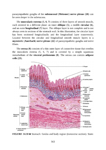
parasympathetic ganglia of the submucosal (Meissner) nerve plexus (20) can

               be seen deeper in the submucosa.

                   The muscularis externa (5, 6, 7) consists of three layers of smooth muscle,
               each oriented in a different plane: an inner oblique (5), a middle circular (6),
               and an outer longitudinal (7) layer. The oblique layer is not complete and is not
               always seen in sections of the stomach wall. In this illustration, the circular layer

               has  been  sectioned  longitudinally  and  the  longitudinal  layer  transversely.
               Located  between  the  circular  and  longitudinal  smooth  muscle  layers  is  a
               myenteric (Auerbach) nerve plexus (22) of parasympathetic ganglia and nerve
               fibers.


                   The serosa (8) consists of a thin outer layer of connective tissue that overlies
               the  muscularis  externa  (5,  6,  7)  and  is  covered  by  a  simple  squamous
               mesothelium  of  the  visceral peritoneum (8).  The  serosa  can  contain  adipose
               cells (23).


               FIGURE 14.10 ■ Stomach: fundus and body region (transverse section). Stain:


                                                          563