
Page 559 - Atlas of Histology with Functional Correlations
P. 559
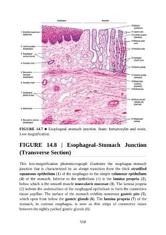
FIGURE 14.7 ■ Esophageal–stomach junction. Stain:  hematoxylin  and  eosin.
               Low magnification.


               FIGURE  14.8  |  Esophageal–Stomach  Junction

               (Transverse Section)


               This  low-magnification  photomicrograph  illustrates  the  esophagus–stomach
               junction  that  is  characterized  by  an  abrupt  transition  from  the  thick  stratified
               squamous epithelium (1) of the esophagus to the simple columnar epithelium
               (4)  of  the  stomach.  Inferior  to  the  epithelium  (1)  is  the  lamina  propria  (2),

               below which is the smooth muscle muscularis mucosae (3). The lamina propria
               (2) indents the undersurface of the esophageal epithelium to form the connective
               tissue papillae. The surface of the stomach exhibits numerous gastric pits (5),
               which open from below the gastric glands (6). The lamina propria (7) of the

               stomach,  in  contrast  esophagus,  is  seen  as  thin  strips  of  connective  tissue
               between the tightly packed gastric glands (6).


                                                          558