
Page 555 - Atlas of Histology with Functional Correlations
P. 555
region.
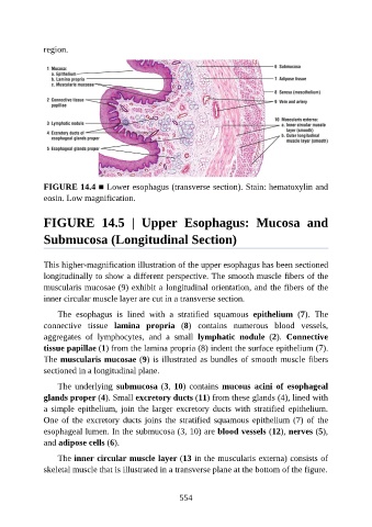
FIGURE 14.4 ■ Lower esophagus (transverse section). Stain: hematoxylin and
eosin. Low magnification.
FIGURE 14.5 | Upper Esophagus: Mucosa and
Submucosa (Longitudinal Section)
This higher-magnification illustration of the upper esophagus has been sectioned
longitudinally to show a different perspective. The smooth muscle fibers of the
muscularis mucosae (9) exhibit a longitudinal orientation, and the fibers of the
inner circular muscle layer are cut in a transverse section.
The esophagus is lined with a stratified squamous epithelium (7). The
connective tissue lamina propria (8) contains numerous blood vessels,
aggregates of lymphocytes, and a small lymphatic nodule (2). Connective
tissue papillae (1) from the lamina propria (8) indent the surface epithelium (7).
The muscularis mucosae (9) is illustrated as bundles of smooth muscle fibers
sectioned in a longitudinal plane.
The underlying submucosa (3, 10) contains mucous acini of esophageal
glands proper (4). Small excretory ducts (11) from these glands (4), lined with
a simple epithelium, join the larger excretory ducts with stratified epithelium.
One of the excretory ducts joins the stratified squamous epithelium (7) of the
esophageal lumen. In the submucosa (3, 10) are blood vessels (12), nerves (5),
and adipose cells (6).
The inner circular muscle layer (13 in the muscularis externa) consists of
skeletal muscle that is illustrated in a transverse plane at the bottom of the figure.
554