
Page 554 - Atlas of Histology with Functional Correlations
P. 554
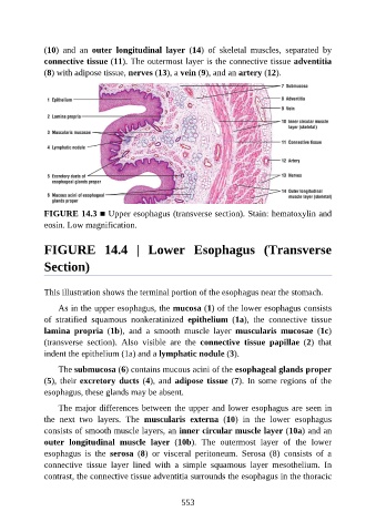
(10)  and  an  outer  longitudinal  layer  (14)  of  skeletal  muscles,  separated  by

               connective tissue (11). The outermost layer is the connective tissue adventitia
               (8) with adipose tissue, nerves (13), a vein (9), and an artery (12).


               FIGURE 14.3 ■ Upper esophagus (transverse section). Stain: hematoxylin and
               eosin. Low magnification.


               FIGURE  14.4  |  Lower  Esophagus  (Transverse


               Section)


               This illustration shows the terminal portion of the esophagus near the stomach.

                   As in the upper esophagus, the mucosa (1) of the lower esophagus consists
               of  stratified  squamous  nonkeratinized  epithelium  (1a),  the  connective  tissue

               lamina  propria  (1b),  and  a  smooth  muscle  layer  muscularis  mucosae  (1c)
               (transverse  section).  Also  visible  are  the  connective  tissue  papillae  (2)  that
               indent the epithelium (1a) and a lymphatic nodule (3).

                   The submucosa (6) contains mucous acini of the esophageal glands proper

               (5), their excretory ducts (4), and adipose tissue (7). In some regions of the
               esophagus, these glands may be absent.

                   The major differences between the upper and lower esophagus are seen in
               the  next  two  layers.  The  muscularis  externa  (10)  in  the  lower  esophagus
               consists of smooth muscle layers, an inner circular muscle layer (10a) and an

               outer  longitudinal  muscle  layer  (10b).  The  outermost  layer  of  the  lower
               esophagus  is  the  serosa  (8)  or  visceral  peritoneum.  Serosa  (8)  consists  of  a
               connective  tissue  layer  lined  with  a  simple  squamous  layer  mesothelium.  In

               contrast, the connective tissue adventitia surrounds the esophagus in the thoracic


                                                          553