
Page 557 - Atlas of Histology with Functional Correlations
P. 557
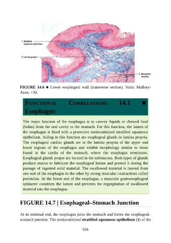
FIGURE  14.6  ■  Lower  esophageal  wall  (transverse  section).  Stain:  Mallory-
               Azan. ×30.


                  FUNCTIONAL                     CORRELATIONS                       14.1            ■


                  Esophagus


                 The  major  function  of  the  esophagus  is  to  convey  liquids  or  chewed  food
                 (bolus) from the oral cavity to the stomach. For this function, the lumen of
                 the esophagus is lined with a protective nonkeratinized stratified squamous

                 epithelium. Aiding in this function are esophageal glands in lamina propria.
                 The  esophageal  cardiac  glands  are  in  the  lamina  propria  of  the  upper  and
                 lower  regions  of  the  esophagus  and  exhibit  morphology  similar  to  those

                 found  in  the  cardia  of  the  stomach,  where  the  esophagus  terminates.
                 Esophageal glands proper are located in the submucosa. Both types of glands
                 produce mucus to lubricate the esophageal lumen and protect it during the
                 passage of ingested solid material. The swallowed material is moved from
                 one end of the esophagus to the other by strong muscular contractions called

                 peristalsis. At the lower end of the esophagus, a muscular gastroesophageal
                 sphincter constricts the lumen and prevents the regurgitation of swallowed
                 material into the esophagus.


               FIGURE 14.7 | Esophageal–Stomach Junction


               At its terminal end, the esophagus joins the stomach and forms the esophageal–
               stomach junction. The nonkeratinized stratified squamous epithelium (1) of the


                                                          556