
Page 556 - Atlas of Histology with Functional Correlations
P. 556
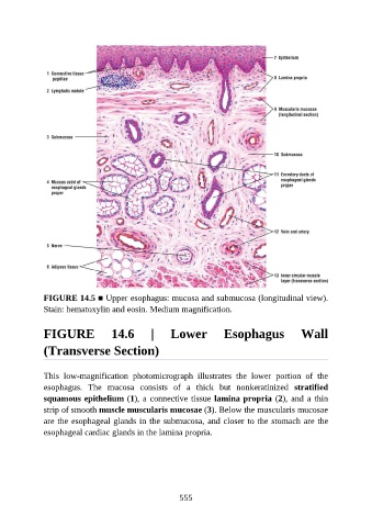
FIGURE 14.5 ■ Upper esophagus: mucosa and submucosa (longitudinal view).
               Stain: hematoxylin and eosin. Medium magnification.


               FIGURE                14.6        |      Lower            Esophagus               Wall

               (Transverse Section)


               This  low-magnification  photomicrograph  illustrates  the  lower  portion  of  the

               esophagus.  The  mucosa  consists  of  a  thick  but  nonkeratinized  stratified
               squamous epithelium (1), a connective tissue lamina propria (2), and a thin
               strip of smooth muscle muscularis mucosae (3). Below the muscularis mucosae
               are the esophageal glands in the submucosa, and closer to the stomach are the

               esophageal cardiac glands in the lamina propria.


                                                          555