
Page 551 - Atlas of Histology with Functional Correlations
P. 551
adventitia) in the wall of the esophagus and their characteristic contents.
The esophageal lumen is lined with a moist, nonkeratinized stratified
squamous epithelium. An empty esophagus exhibits numerous but temporary
longitudinal folds of mucosa in its lumen that are due to the contractions of the
esophageal muscles. The wall of the esophagus contains two types of glands that
secrete mucus; however, they are located in different parts of the organ. In the
lamina propria of the proximal and distal parts of the esophagus near the
stomach are the esophageal cardiac glands because they resemble the mucous
glands located in the cardiac region of the stomach. In the submucosa are the
esophageal glands proper that are scattered along the entire length of the
esophagus. The mucus from these glands lubricates the lumen of the esophagus,
protects the mucosa, and facilitates smooth passage of food material (bolus)
through the esophagus to the stomach.
The outer wall of the esophagus, the muscularis externa, contains both
skeletal and smooth muscles fibers. In the upper third of the esophagus, both
layers of the muscularis externa contain striated skeletal muscle fibers. In the
middle third of the esophagus, the muscularis externa contains a mixture of both
skeletal and smooth muscle fibers, whereas in the lower third of the esophagus,
both layers are smooth muscle fibers (see Fig. 14.1).
Supplemental micrographic images are available at
www.thePoint.com/Eroschenko13e under Digestive System Part II:
Esophagus and Stomach.
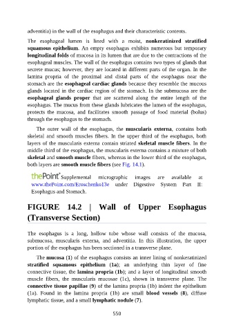
FIGURE 14.2 | Wall of Upper Esophagus
(Transverse Section)
The esophagus is a long, hollow tube whose wall consists of the mucosa,
submucosa, muscularis externa, and adventitia. In this illustration, the upper
portion of the esophagus has been sectioned in a transverse plane.
The mucosa (1) of the esophagus consists an inner lining of nonkeratinized
stratified squamous epithelium (1a); an underlying thin layer of fine
connective tissue, the lamina propria (1b); and a layer of longitudinal smooth
muscle fibers, the muscularis mucosae (1c), shown in transverse plane. The
connective tissue papillae (9) of the lamina propria (1b) indent the epithelium
(1a). Found in the lamina propria (1b) are small blood vessels (8), diffuse
lymphatic tissue, and a small lymphatic nodule (7).
550